A multidisciplinary research team from the University of Macau (UM) recently achieved a significant breakthrough in drug susceptibility test for cancer cell lines and human primary cancers by combining their expertise in precision medicine, microfluidic chip, and image processing. The screening method shortened the analysis process to within 24 hours, which was made possible by a cutting-edge, state-of-the-art technology developed in-house that merges biology and electronics, which laid a foundation for evidence-based drug susceptibility tests for precision cancer therapy. The work was recently published in the open access journal Scientific Reports under the Nature Publishing Group.
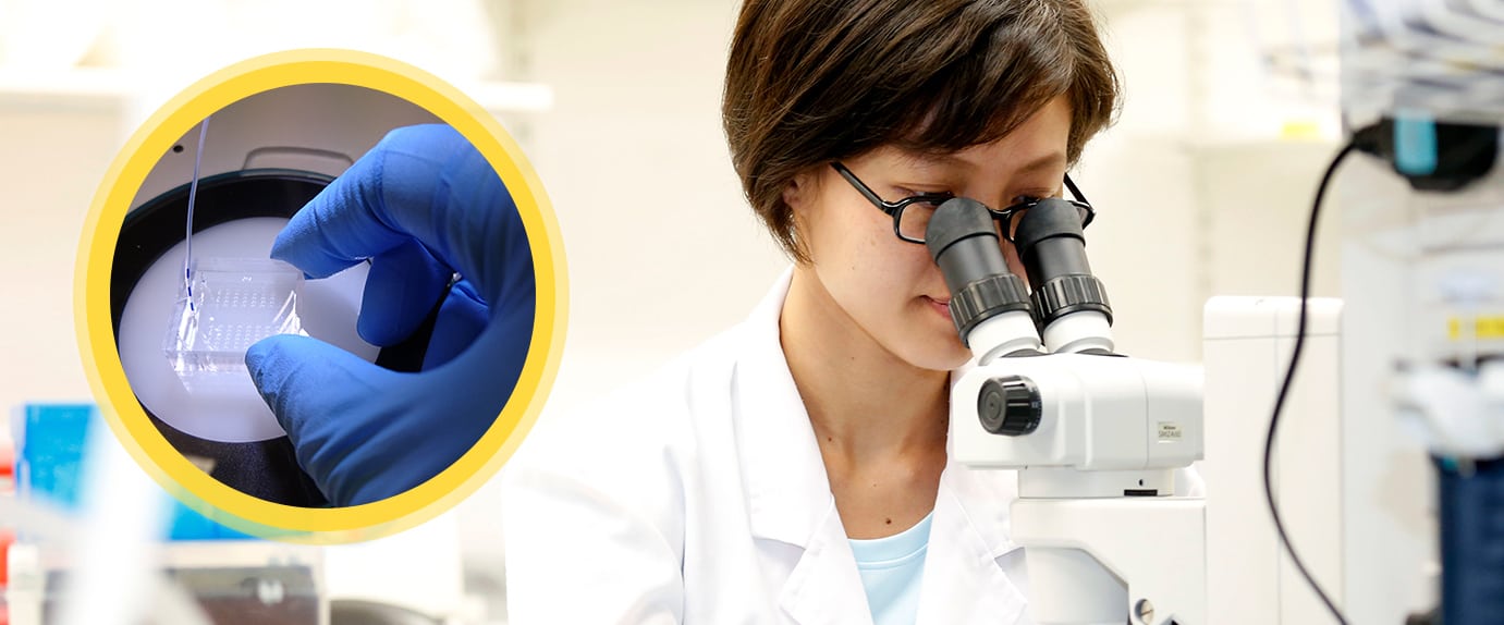
This project was mainly contributed by Ada Wong Hang Heng, UM Macao Fellow of the Faculty of Health Sciences (FHS), under the leadership of FHS Dean Chuxia Deng. Wong performed all the experiments and applied a microchip to drug screening for cells and human primary tumors. The paper entitled ‘Drug Screening of Cancer Cell Lines and Human Primary Tumors Using Droplet Microfluidics’ was published in the Scientific Reports. In this study, UM researchers tested the susceptibility of cells dissociated from cancer tissues to different drugs on a microfluidic chip developed by the State Key Laboratory of Analog and Mixed Signal VLSI (AMSV). Exploitation of an artificial intelligence-based image processing technology developed by the Faculty of Science and Technology (FST) facilitated the observation of single cell viability in each microfluidic droplet post-treatment for the purpose of drug screening, and achieved results within 24 hours. The screening method laid a foundation for evidence-based drug susceptibility tests for precision cancer therapy. The resolution and statistical capability of this assay opened the door to studying single cell differential drug response within heterogeneous cancers.
This study used an improved microchip measuring 2.4 x 2.4 cm in dimension. The cost of chip fabrication was low. 16,000 cells were applied for every drug screening condition. The current protocol succeeded in parallel screening of a maximum of nine drugs per chip, whereas the team’s target throughput is 96 drugs per chip after process automation. Reported primary cancer cell screening platforms either only allowed screening of a few drugs, or required amplified culture to achieve large-scale drug screening, which took at least several weeks to complete. The new microchip enabled drug susceptibility test of single cells in microfluidic droplets from a small sample size, which, in combination with image processing, expedited the drug screening process. This assay gave results within 24 hours.
The microfluidic chip used in the study was developed by researchers from the State Key Laboratory of Analog and Mixed Signal VLSI (AMSV), namely AMSV Director Rui Martins, Associate Professor Mak Pui In, and Assistant Professor Jia Yanwei. The automatic image-processing method was developed by Prof Wong Pak Kin and Prof Vong Chi Man from the FST. This project was funded by the Macau Science and Technology Development Fund (FDCT) and received support from Kiang Wu Hospital.
For Chinese version, please visit here.